Aim: The aim of this study is to report the computed tomography characteristics of the degeneration of Crohn's disease. Patient: We report the observation of an 18-year-old girl admitted to the internal medicine department of the CHU Point G for diffuse abdominal pain predominantly in the right iliac fossa with diarrhea that has progressed since childhood but poorly followed. In whom the abdominal CT scan and abdominopelvic ultrasound performed revealed irregular vascular thickening of the wall of the terminal ileum and the cecum. At the end of these examinations, the diagnostic hypothesis of atypical Crohn's disease was adopted. Histology of the transverse colon specimen showed fragments of mucosa with an edematous chorion and an inflammatory infiltrate of omental and gigantocellular granuloma. On the other hand, that of the part of the ileocecal region showed a persistence of mucosecretion and the presence of columnar cells with an oval nucleus not exceeding 3/4 of the height of the epithelium and a little marked polymorphism in connection with neoplasia. certain low grade intraepithelial colic. This examination confirmed the diagnosis of carcinomatous degeneration of ileo-caeco-colic Crohn's disease. The clinical course at 6 months was favorable. However, the irregular thickening of the wall of the terminal ileum and the cecum is similar to the CT scan. Conclusion: Degeneration of Crohn's disease is rare. Imaging is involved in all phases of Crohn's disease. She makes the initial diagnosis, she specifies the topographical assessment of the lesions, she guides the treatment, she draws up the extension assessment during an evolutionary outbreak and degeneration.
This is an Open Access article, distributed under the terms of the Creative Commons Attribution 4.0 International License (http://creativecommons.org/licenses/by/4.0/), which permits unrestricted use, distribution and reproduction in any medium or format, provided the original work is properly cited.
Crohn's disease is a chronic intestinal inflammatory disease of the gastrointestinal tract which most often affects young adults characterized by elective involvement of the terminal ileum in 80% of cases and also the colon in 50% of cases
[1]
Herlinger H, Caroline DF. Crohn's disease of the small bowel. In: Gore RM, Levine MS, editors. Textbook of gastrointestinal radiology. Philadelphia: W. B. Saunders; 2000. p. 726-45.
[1]
.
The chronic course of the disease is characterized by flare-ups interspersed with phases of remission. This variable behavior has led to a classification into different types including active inflammatory forms, stenosing forms and complicated forms. Among these potential complications, carcinomatous degeneration, although rare, constitutes both a diagnostic and therapeutic problem. Although this development seems avoidable by endoscopic monitoring and radiological explorations, incidental diagnoses remain possible, including at advanced stages
[2]
Lerebours E., Michel P. (1995) Crohn's disease. In: Bouvenot G., Devulder B., Guillevin L., Queneau P., Schaeffer A. Medical pathology, Gastroenterology, Hepatology, Hematology. Vol. 4. Paris: Masson, p. 135.136.
[2]
.
The risk of degeneration is similar between Crohn's disease and ulcerative colitis: in patients operated on for chronic colonic or rectal inflammatory bowel disease, the occurrence of colorectal cancer does not differ depending on the type of disease. However, the prognosis for colorectal cancer arising from Crohn's disease seems more poor
[3]
Svrcek M, Cosnes J, Beaugerie L, Parc R, Bennis M, Tiret E, et al. Colorectal neoplasia in Crohn’s colitis: a retrospective comparative study with ulcerative colitis. Histopathology. 2007 Apr; 50(5): 574-83.
[3]
.
Digestive exploration using medical imaging techniques is essential to establish the diagnosis of Crohn's disease. Endoscopy is always the technique used first
[4]
Beau P., Gay G., Arpurt J. P., Boustière C. et al. (2004) Role of endoscopy in the assessment of Crohn's disease. In: Recommendation of the French Society of Digestive Endoscopy. [online]. (Accessed November 17, 2011). Available at:
The advent of cross-sectional imaging has modified the diagnostic semiology by improving the assessment of extension and revealing complications early.
The radiological appearance and evolution over time are variable, marked by numerous flare-ups and various complications (fistulas, mesenteric abscess and degeneration, etc.)
The management of this degeneration is thus a source of difficulties, both in its diagnosis, in the individual evaluation of the prognosis associated with it, and also in the therapeutic strategy, particularly surgical.
In Africa in general and in Mali in particular, to our knowledge very few cases are reported in the literature, hence the interest in studying this case.
We initiated this work with the aim of reporting the CT characteristics of the degeneration of Crohn's disease.
The objective of this work is to report the CT characteristics of the degeneration of Crohn's disease at Point G University Hospital.
2. Observation
This is an 18-year-old girl seen in the internal medicine department of Point G University Hospital for Crohn's disease. She complained intermittently of diffuse abdominal pain predominating in the right iliac fossa with diarrhea and fever since the age of 5 years, poorly followed. She received treatments based on Ibuprofen 400mg and Smecta sachet. She was admitted to the emergency room of the Point G University Hospital for acute abdominal pain lasting for 2 days for which a Fast Echo was carried out and wrongly concluded to be appendicitis. In view of this, a thorough pre-therapeutic assessment was requested showing an inflammatory syndrome with CRP. at 6 times normal, i.e. 37ng/l and an ESR of 16mm/s in the 1st hour and 44mm/s in the 2nd hour. The blood count was normal.
Clinically, on admission: during the clinical examination, the patient was conscious and active; emaciated with a BMI equal to 11 (i.e. height equal to 176cm and a weight of 37kgs with a T° of 37.5°C), the integuments and mucous membranes were well colored.
On physical examination, inspection revealed a flexible abdomen participating in breathing.
On palpation we noted a slight pain in the MC Burney point radiating to the umbilicus.
On cardiovascular auscultation, the heart rate was 83 beats/min, the blood pressure was 123/75 mmHg in both arms.
In terms of morphological examination, an abdominal scan is requested. This examination was carried out according to the classic protocol: a first acquisition without injection followed by an intravenous injection of 80ml of Omnipaque at 2.5ml/s then acquisition in the arterial phase at 25S and in the portal phase at 70S.
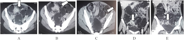
This examination allowed us to demonstrate a dedifferentiated parietal thickening of the distal part of the terminal ileum extended over 6cm also involving the cecal wall. This thickening is irregular and stenosing with a discreet infiltration of the peri-lesional fat. The thickened digestive walls were enhanced homogeneously after injection of contrast product.
We also noted small areas of thickening 7mm in places on the transverse colon spaced from large normal colic areas.
The easily spotted appendix was normal in appearance, the diagnostic hypothesis of appendicitis was ruled out and that of atypical Crohn's disease was retained.
Ileo-colonoscopy carried out under propopol 200mg. The colon was examined up to the cecum, the colonic mucosa presented healthy areas and pathological areas. In the pathological areas there were wounds in the form of rail ulcerations, stenosing in places, the largest of which measured approximately 1.5cm in diameter covered in fibrin in the cecum region. Ileoscopy could not be performed due to the probe not passing through. However, two biopsies were taken, one on the terminal ileum and the other on the transverse colon, particularly on the pathological area of the transverse colon. This examination made it possible to establish the diagnostic hypothesis of chronic inflammatory intestinal disease associated with a tumor of the transverse colon.
The histology of the transverse colon showed fragments of mucosa, the chorion of which was edematous and the site of an inflammatory infiltrate of omental and gigantocellular granuloma. On the other hand, that of the piece from the ileo-caecal region showed a persistence of mucosecretion and the presence of cylindrical cells with an oval nucleus not exceeding 3/4 of the height of the epithelium and a little marked polymorphism in relation to neoplasia certain low grade colonic intraepithelial.
Histology confirmed the diagnosis of carcinomatous degeneration of ileocecocolic Crohn's disease.
Our patient was put on treatment based on corticosteroids (SOLUPRED effervescent tablet at 10 mg in the morning), amino-salicylated derivatives (PENTASA sachets 1000).
Surgical treatment was proposed and refused by the patient and her parents.
The CT scan at 6 months of treatment showed stability of the thickening of the terminal ileum and a regression of other colonic thickenings.
Figure 1. Axial (A, B and C) and coronal (D and E) CT sections after injection of contrast product showing enhancement of the thickening of the wall of the terminal ileum and the cecum (arrow).
3. Discussion
Crohn's disease seems to be favored by genetic, vascular, infectious, dietary and psychological factors
[2]
Lerebours E., Michel P. (1995) Crohn's disease. In: Bouvenot G., Devulder B., Guillevin L., Queneau P., Schaeffer A. Medical pathology, Gastroenterology, Hepatology, Hematology. Vol. 4. Paris: Masson, p. 135.136.
[2]
. Our patient was an 18-year-old girl with no elucidated genetic predisposition factors.
The disease is characterized by the alternation of flare-ups of varying intensity and periods of remission of varying duration, and more or less complete
[5]
HAS, Haute Autorité de Santé: (2008) ALD 24 guide “Crohn’s Disease” [online]. p. 4. (Accessed September 21, 2011). Available at:
Digestive manifestations are numerous, associated or isolated, depending on the severity and location of the lesions. They are indicative of Crohn's disease in the majority of cases
[6]
Lerebours E., Michel P. (1995) Crohn's disease. In: Bouvenot G., Devulder B., Guillevin L., Queneau P., Schaeffer A. Medical pathology, Gastroenterology, Hepatology, Hematology. Vol. 4. Paris: Masson, p. 137.
[6]
. Crohn's disease can be associated with inflammatory manifestations affecting other organs. We are talking about extradigestive manifestations causing lesions, most often of the joints, skin and eyes. Our patient did not present any extradigestive signs.
In our case, these two symptoms were the reasons for consultation of our patient who was characterized by appendicular syndrome type pain, with throbbing and fixed pain, associated with meteorism. They are relieved by the emission of gas or stools.
A deterioration in general condition often accompanies outbreaks of the disease
[7]
AFA: François Aupetit Association, overcoming Crohn's disease and ulcerative colitis. (June 2008) What causes Crohn's disease? [Online]. (Accessed September 21, 2011). Available at:
. Our patient only had episodes of fever and almost constant weight loss due to diarrhea with a body mass index (BMI) of 11.
Age at diagnosis and duration of progression can be considered as a major risk factor for degeneration. This has been mentioned in other studies in the general population, finding a relative risk of 4 in patients diagnosed before the age of 15
[8]
Ekbom A, Helmick C, Zack M, Adami HO. Ulcerative colitis and colorectal cancer. A population-based study. N Engl J Med. 1990 Nov 1; 323(18): 1228-33.
[8]
. This is, however, contradicted by other works. This factor is also possibly linked to the duration of evolution and its independent nature remains uncertain. Which is fully consistent with our study framework.
A family history of colorectal cancer has been found in several large studies as a risk factor for degeneration
[9]
Askling J, Dickman PW, Karlén P, Broström O, Lapidus A, Löfberg R, et al. Family history as a risk factor for colorectal cancer in inflammatory bowel disease. Gastroenterology. 2001 May; 120(6): 1356-62.
[9]
. We did not highlight this criterion, possibly taking into account a single case study.
The male sex could be mentioned as statistically associated with degeneration, in particular by Soderlund et al.
[10]
Söderlund S, Granath F, Broström O, Karlén P, Löfberg R, Ekbom A, et al. Inflammatory bowel disease confers a lower risk of colorectal cancer to females than to males. Gastroenterology. 2010 May; 138(5): 1697-703.
[10]
. This is contrary to our case.
Reflux ileitis was mentioned by Heuschen as a risk factor for degeneration, not found in several other studies, this factor was not elucidated in our study
[11]
Heuschen UA, Hinz U, Allemeyer EH, Stern J, Lucas M, Autschbach F, et al. Backwash ileitis is strongly associated with colorectal carcinoma in ulcerative colitis. Gastroenterology. 2001 Mar; 120(4): 841-7.
[11]
.
The existence of dysplasia, a precancerous condition, constitutes a marker of the risk of degeneration. This is consistent with our patient who presented dysphasia in the ileo-caecal region and inflammatory cells in the transverse colon.
The initial assessment, patient care and making a definitive diagnosis involve a multidisciplinary team made up of the attending physician, hepato-gastroenterologist, biologists, radiologists, pathologists and, if necessary, other specialists such as the rheumatologist, dermatologist and ophthalmologist. The diagnosis is actually based on a range of clinical and para-clinical arguments involving biology and medical imaging
[12]
HAS, Haute Autorité de Santé: (2008) ALD 24 guide “Crohn’s disease”. p. 5-6. [Online]. (Accessed November 15, 2011). Available at:
In our patient the biology showed an increase in the CRP level to 37 mg/l, polynuclear neutrophils to 8658/mm3, an acceleration of the sedimentation rate at the 1st hour 16mm and 2nd hour 44mm. The hemoglobin level was of 12g/100ml.
On imaging, the abdominopelvic ultrasound revealed a dedifferentiated thickening of the wall of the digestive tract at the level of the right iliac fossa with an inflammatory appearance.
The abdominal scan was carried out according to the colon scanner protocol, helical acquisition without injection of the iodinated contrast product before an evacuating enema of the colon with water, then colonic filling with 1.5 ml of water followed by the injection of the product contrast and acquisition in the arterial and protal phases.
This examination allowed us to identify two thickenings, the first irregular and circumferential parietal thickening of the last ileal loop measuring 15mm thick by 66mm high. This thickening extended to the proximal part of the cecum.
The second thickening which was non-stenotic circumferential with regular contours at the level of the middle portion of the transverse colon measured at 16mm thickness.
Following these two radiological examinations, the diagnostic hypothesis of ileocecal Crohn's disease and atypical transverse colon was raised.
The total vaginal ileocolonoscopy of our patient with biopsy in diseased and healthy areas is the reference examination to confirm the diagnosis and to evaluate the topography and severity of Crohn's disease.
In endoscopy, we observe the high frequency of flat and stenosing lesions compared to budding lesions. Gumaste et al found a very high risk of degeneration and underlined the need to detect these stenoses, often associated with more advanced lesions with an infiltrative nature
[5]
HAS, Haute Autorité de Santé: (2008) ALD 24 guide “Crohn’s Disease” [online]. p. 4. (Accessed September 21, 2011). Available at:
. This therefore confirms the “risky” nature of stenoses, which must be detected. Other lesions, non-stenosing, may be found. In our case, unlike several other series, the histology of the ileocecal part presented fragments of mucosa whose chorion is edematous and the site of an inflammatory infiltrate of omental and gigantocellular granuloma. On the other hand, that of the piece from the transverse colon region showed a persistence of mucosecretion and the presence of cylindrical cells with an oval nucleus not exceeding 3/4 of the height of the epithelium and a little marked polymorphism in relation to neoplasia certain low grade colonic intraepithelial.
4. Conclusion
Degeneration of Crohn's disease is rare. Several risk factors make it possible to estimate its occurrence, in addition to the recommended endoscopic screening.
Imaging is involved in all phases of Crohn's disease. She makes the initial diagnosis, she specifies the topographical assessment of the lesions, she directs the treatment and evaluates the causes of ineffective treatment and finally, she develops the assessment of extension during a progressive flare-up.
Abbreviations
CRP
C-reactive Protein
BMI
Body Mass Index
CHU
University Hospital Center
VS
Sedimentation Speed
S
Second
T°
Temperature
CD
Crohn's Disease
Author Contributions
All authors contributed to data acquisition, data analysis and interpretation, and writing of the article.
Conflicts of Interest
The authors declare no conflicts of interest.
References
[1]
Herlinger H, Caroline DF. Crohn's disease of the small bowel. In: Gore RM, Levine MS, editors. Textbook of gastrointestinal radiology. Philadelphia: W. B. Saunders; 2000. p. 726-45.
[2]
Lerebours E., Michel P. (1995) Crohn's disease. In: Bouvenot G., Devulder B., Guillevin L., Queneau P., Schaeffer A. Medical pathology, Gastroenterology, Hepatology, Hematology. Vol. 4. Paris: Masson, p. 135.136.
[3]
Svrcek M, Cosnes J, Beaugerie L, Parc R, Bennis M, Tiret E, et al. Colorectal neoplasia in Crohn’s colitis: a retrospective comparative study with ulcerative colitis. Histopathology. 2007 Apr; 50(5): 574-83.
[4]
Beau P., Gay G., Arpurt J. P., Boustière C. et al. (2004) Role of endoscopy in the assessment of Crohn's disease. In: Recommendation of the French Society of Digestive Endoscopy. [online]. (Accessed November 17, 2011). Available at:
Lerebours E., Michel P. (1995) Crohn's disease. In: Bouvenot G., Devulder B., Guillevin L., Queneau P., Schaeffer A. Medical pathology, Gastroenterology, Hepatology, Hematology. Vol. 4. Paris: Masson, p. 137.
[7]
AFA: François Aupetit Association, overcoming Crohn's disease and ulcerative colitis. (June 2008) What causes Crohn's disease? [Online]. (Accessed September 21, 2011). Available at:
Ekbom A, Helmick C, Zack M, Adami HO. Ulcerative colitis and colorectal cancer. A population-based study. N Engl J Med. 1990 Nov 1; 323(18): 1228-33.
[9]
Askling J, Dickman PW, Karlén P, Broström O, Lapidus A, Löfberg R, et al. Family history as a risk factor for colorectal cancer in inflammatory bowel disease. Gastroenterology. 2001 May; 120(6): 1356-62.
[10]
Söderlund S, Granath F, Broström O, Karlén P, Löfberg R, Ekbom A, et al. Inflammatory bowel disease confers a lower risk of colorectal cancer to females than to males. Gastroenterology. 2010 May; 138(5): 1697-703.
[11]
Heuschen UA, Hinz U, Allemeyer EH, Stern J, Lucas M, Autschbach F, et al. Backwash ileitis is strongly associated with colorectal carcinoma in ulcerative colitis. Gastroenterology. 2001 Mar; 120(4): 841-7.
[12]
HAS, Haute Autorité de Santé: (2008) ALD 24 guide “Crohn’s disease”. p. 5-6. [Online]. (Accessed November 15, 2011). Available at:
Mamoudou, C., Aminata, S., Toumin, C., Abdoulaye, K., Moussa, K., et al. (2025). Carcinomatous Degeneration of Crohn's Disease About a Case at G-Point CHU / Mali. International Journal of Medical Case Reports, 4(1), 17-20. https://doi.org/10.11648/j.ijmcr.20250401.13
Mamoudou, C.; Aminata, S.; Toumin, C.; Abdoulaye, K.; Moussa, K., et al. Carcinomatous Degeneration of Crohn's Disease About a Case at G-Point CHU / Mali. Int. J. Med. Case Rep.2025, 4(1), 17-20. doi: 10.11648/j.ijmcr.20250401.13
Mamoudou C, Aminata S, Toumin C, Abdoulaye K, Moussa K, et al. Carcinomatous Degeneration of Crohn's Disease About a Case at G-Point CHU / Mali. Int J Med Case Rep. 2025;4(1):17-20. doi: 10.11648/j.ijmcr.20250401.13
@article{10.11648/j.ijmcr.20250401.13,
  author = {Camara Mamoudou and Sakho Aminata and Camara Toumin and Koné Abdoulaye and Konaté Moussa and Koné Youssouf and Sidibé Kassim and Coulibaly Youlouza and Sidibé Siaka},
  title = {Carcinomatous Degeneration of Crohn's Disease About a Case at G-Point CHU / Mali
},
  journal = {International Journal of Medical Case Reports},
  volume = {4},
  number = {1},
  pages = {17-20},
  doi = {10.11648/j.ijmcr.20250401.13},
  url = {https://doi.org/10.11648/j.ijmcr.20250401.13},
  eprint = {https://article.sciencepublishinggroup.com/pdf/10.11648.j.ijmcr.20250401.13},
  abstract = {Aim: The aim of this study is to report the computed tomography characteristics of the degeneration of Crohn's disease. Patient: We report the observation of an 18-year-old girl admitted to the internal medicine department of the CHU Point G for diffuse abdominal pain predominantly in the right iliac fossa with diarrhea that has progressed since childhood but poorly followed. In whom the abdominal CT scan and abdominopelvic ultrasound performed revealed irregular vascular thickening of the wall of the terminal ileum and the cecum. At the end of these examinations, the diagnostic hypothesis of atypical Crohn's disease was adopted. Histology of the transverse colon specimen showed fragments of mucosa with an edematous chorion and an inflammatory infiltrate of omental and gigantocellular granuloma. On the other hand, that of the part of the ileocecal region showed a persistence of mucosecretion and the presence of columnar cells with an oval nucleus not exceeding 3/4 of the height of the epithelium and a little marked polymorphism in connection with neoplasia. certain low grade intraepithelial colic. This examination confirmed the diagnosis of carcinomatous degeneration of ileo-caeco-colic Crohn's disease. The clinical course at 6 months was favorable. However, the irregular thickening of the wall of the terminal ileum and the cecum is similar to the CT scan. Conclusion: Degeneration of Crohn's disease is rare. Imaging is involved in all phases of Crohn's disease. She makes the initial diagnosis, she specifies the topographical assessment of the lesions, she guides the treatment, she draws up the extension assessment during an evolutionary outbreak and degeneration.
},
 year = {2025}
}
TY  - JOUR
T1  - Carcinomatous Degeneration of Crohn's Disease About a Case at G-Point CHU / Mali

AU  - Camara Mamoudou
AU  - Sakho Aminata
AU  - Camara Toumin
AU  - Koné Abdoulaye
AU  - Konaté Moussa
AU  - Koné Youssouf
AU  - Sidibé Kassim
AU  - Coulibaly Youlouza
AU  - Sidibé Siaka
Y1  - 2025/02/21
PY  - 2025
N1  - https://doi.org/10.11648/j.ijmcr.20250401.13
DO  - 10.11648/j.ijmcr.20250401.13
T2  - International Journal of Medical Case Reports
JF  - International Journal of Medical Case Reports
JO  - International Journal of Medical Case Reports
SP  - 17
EP  - 20
PB  - Science Publishing Group
SN  - 2994-7049
UR  - https://doi.org/10.11648/j.ijmcr.20250401.13
AB  - Aim: The aim of this study is to report the computed tomography characteristics of the degeneration of Crohn's disease. Patient: We report the observation of an 18-year-old girl admitted to the internal medicine department of the CHU Point G for diffuse abdominal pain predominantly in the right iliac fossa with diarrhea that has progressed since childhood but poorly followed. In whom the abdominal CT scan and abdominopelvic ultrasound performed revealed irregular vascular thickening of the wall of the terminal ileum and the cecum. At the end of these examinations, the diagnostic hypothesis of atypical Crohn's disease was adopted. Histology of the transverse colon specimen showed fragments of mucosa with an edematous chorion and an inflammatory infiltrate of omental and gigantocellular granuloma. On the other hand, that of the part of the ileocecal region showed a persistence of mucosecretion and the presence of columnar cells with an oval nucleus not exceeding 3/4 of the height of the epithelium and a little marked polymorphism in connection with neoplasia. certain low grade intraepithelial colic. This examination confirmed the diagnosis of carcinomatous degeneration of ileo-caeco-colic Crohn's disease. The clinical course at 6 months was favorable. However, the irregular thickening of the wall of the terminal ileum and the cecum is similar to the CT scan. Conclusion: Degeneration of Crohn's disease is rare. Imaging is involved in all phases of Crohn's disease. She makes the initial diagnosis, she specifies the topographical assessment of the lesions, she guides the treatment, she draws up the extension assessment during an evolutionary outbreak and degeneration.

VL  - 4
IS  - 1
ER  -
Faculty of Medicine, Health Sciences and Techniques, Gamal Abdel Nasser University of Conakry, Conakry, Guinea; Medical Imaging Department, CHU Point G, Bamako, Mali
Mamoudou, C., Aminata, S., Toumin, C., Abdoulaye, K., Moussa, K., et al. (2025). Carcinomatous Degeneration of Crohn's Disease About a Case at G-Point CHU / Mali. International Journal of Medical Case Reports, 4(1), 17-20. https://doi.org/10.11648/j.ijmcr.20250401.13
Mamoudou, C.; Aminata, S.; Toumin, C.; Abdoulaye, K.; Moussa, K., et al. Carcinomatous Degeneration of Crohn's Disease About a Case at G-Point CHU / Mali. Int. J. Med. Case Rep.2025, 4(1), 17-20. doi: 10.11648/j.ijmcr.20250401.13
Mamoudou C, Aminata S, Toumin C, Abdoulaye K, Moussa K, et al. Carcinomatous Degeneration of Crohn's Disease About a Case at G-Point CHU / Mali. Int J Med Case Rep. 2025;4(1):17-20. doi: 10.11648/j.ijmcr.20250401.13
@article{10.11648/j.ijmcr.20250401.13,
  author = {Camara Mamoudou and Sakho Aminata and Camara Toumin and Koné Abdoulaye and Konaté Moussa and Koné Youssouf and Sidibé Kassim and Coulibaly Youlouza and Sidibé Siaka},
  title = {Carcinomatous Degeneration of Crohn's Disease About a Case at G-Point CHU / Mali
},
  journal = {International Journal of Medical Case Reports},
  volume = {4},
  number = {1},
  pages = {17-20},
  doi = {10.11648/j.ijmcr.20250401.13},
  url = {https://doi.org/10.11648/j.ijmcr.20250401.13},
  eprint = {https://article.sciencepublishinggroup.com/pdf/10.11648.j.ijmcr.20250401.13},
  abstract = {Aim: The aim of this study is to report the computed tomography characteristics of the degeneration of Crohn's disease. Patient: We report the observation of an 18-year-old girl admitted to the internal medicine department of the CHU Point G for diffuse abdominal pain predominantly in the right iliac fossa with diarrhea that has progressed since childhood but poorly followed. In whom the abdominal CT scan and abdominopelvic ultrasound performed revealed irregular vascular thickening of the wall of the terminal ileum and the cecum. At the end of these examinations, the diagnostic hypothesis of atypical Crohn's disease was adopted. Histology of the transverse colon specimen showed fragments of mucosa with an edematous chorion and an inflammatory infiltrate of omental and gigantocellular granuloma. On the other hand, that of the part of the ileocecal region showed a persistence of mucosecretion and the presence of columnar cells with an oval nucleus not exceeding 3/4 of the height of the epithelium and a little marked polymorphism in connection with neoplasia. certain low grade intraepithelial colic. This examination confirmed the diagnosis of carcinomatous degeneration of ileo-caeco-colic Crohn's disease. The clinical course at 6 months was favorable. However, the irregular thickening of the wall of the terminal ileum and the cecum is similar to the CT scan. Conclusion: Degeneration of Crohn's disease is rare. Imaging is involved in all phases of Crohn's disease. She makes the initial diagnosis, she specifies the topographical assessment of the lesions, she guides the treatment, she draws up the extension assessment during an evolutionary outbreak and degeneration.
},
 year = {2025}
}
TY  - JOUR
T1  - Carcinomatous Degeneration of Crohn's Disease About a Case at G-Point CHU / Mali

AU  - Camara Mamoudou
AU  - Sakho Aminata
AU  - Camara Toumin
AU  - Koné Abdoulaye
AU  - Konaté Moussa
AU  - Koné Youssouf
AU  - Sidibé Kassim
AU  - Coulibaly Youlouza
AU  - Sidibé Siaka
Y1  - 2025/02/21
PY  - 2025
N1  - https://doi.org/10.11648/j.ijmcr.20250401.13
DO  - 10.11648/j.ijmcr.20250401.13
T2  - International Journal of Medical Case Reports
JF  - International Journal of Medical Case Reports
JO  - International Journal of Medical Case Reports
SP  - 17
EP  - 20
PB  - Science Publishing Group
SN  - 2994-7049
UR  - https://doi.org/10.11648/j.ijmcr.20250401.13
AB  - Aim: The aim of this study is to report the computed tomography characteristics of the degeneration of Crohn's disease. Patient: We report the observation of an 18-year-old girl admitted to the internal medicine department of the CHU Point G for diffuse abdominal pain predominantly in the right iliac fossa with diarrhea that has progressed since childhood but poorly followed. In whom the abdominal CT scan and abdominopelvic ultrasound performed revealed irregular vascular thickening of the wall of the terminal ileum and the cecum. At the end of these examinations, the diagnostic hypothesis of atypical Crohn's disease was adopted. Histology of the transverse colon specimen showed fragments of mucosa with an edematous chorion and an inflammatory infiltrate of omental and gigantocellular granuloma. On the other hand, that of the part of the ileocecal region showed a persistence of mucosecretion and the presence of columnar cells with an oval nucleus not exceeding 3/4 of the height of the epithelium and a little marked polymorphism in connection with neoplasia. certain low grade intraepithelial colic. This examination confirmed the diagnosis of carcinomatous degeneration of ileo-caeco-colic Crohn's disease. The clinical course at 6 months was favorable. However, the irregular thickening of the wall of the terminal ileum and the cecum is similar to the CT scan. Conclusion: Degeneration of Crohn's disease is rare. Imaging is involved in all phases of Crohn's disease. She makes the initial diagnosis, she specifies the topographical assessment of the lesions, she guides the treatment, she draws up the extension assessment during an evolutionary outbreak and degeneration.

VL  - 4
IS  - 1
ER  -


 Share This Article
 Twitter
 Linked In
 Facebook
 Copy
Copy Download